| Sign In | Join Free | My benadorassociates.com |
|
| Sign In | Join Free | My benadorassociates.com |
|
| Categories | Digital Fundus Camera |
|---|---|
| Place of Origin: | China |
| Brand Name: | BIO |
| Model Number: | CRO PLUS |
| MOQ: | 1 unit |
| Delivery Time: | 7 working - days since getting full payment |
| Payment Terms: | Credit card, Paypal, T/T |
| Supply Ability: | 100 units / month |
| Packaging Details: | Carton (75*45*60cm) 15kgs |
| Certification: | CE |
| Model: | CRO PLUS |
| Imaging mode: | FFA + ICGA + AF + RF + NIR + MCOLOR |
| Optical scanning lens: | Optical zoom 100°/60°/30° |
| Fundus imaging FOV: | 160° |
| Light source: | Solid-State LASER |
| Frame frequency: | 16 fps |
| Fundus tracking: | Averaging Real Time Tracking |
| Minimum pupil size: | 2 mm |
| Lesion resolution: | 5 um |
| Video/image output: | Support Avi Jpg Tiff video or image formats |
| Company Info. |
| Wuxi Biomedical Technology Co., Ltd. |
| Verified Supplier |
| View Contact Details |
| Product List |
Ultra-wide Field Laser Scanning Retina Angiograph Digital Ophthalmic Equipment With Optical Zoom 100°/60°/30°
Product Overview
China Leading And Domestic Initiative Confocal Laser Scanning Technology, to Realize Fundus Reflex And Capillary
fluorescence Imaging.
Feature
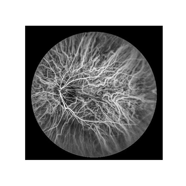
| 1 |  | Fundus Fluorescein Angiography(FFA): Retinal circulatory change | 1) ”Golden standard” to judge retina disease 2) To reflect the physiological pathology of retinal blood vessels to capillaries 3) To comprehensively inspect diseases undetected in normal fundus examination |
| 2 |  | Indocyanine Green Angiography(ICGA) Choroid vessels and RPE lesions | 1) Golden standard for PVC diagnosis 2) To reveal the details of the choroid cycle 3) Supplement to FFA,to find latent CNV undetected on FFA 4) Mainly to reflect blood vessels condition in early and middle stage, in late stage to reflect the form and function of RPE cells |
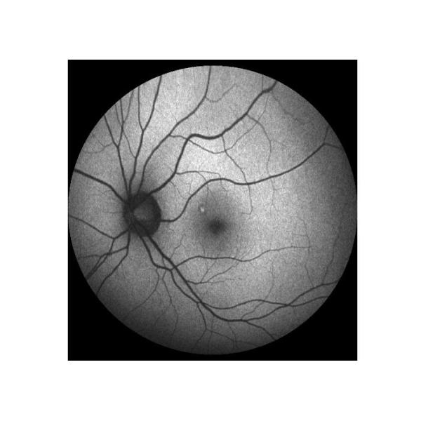
| 3 |  | Infrared(NIR) | 1) To clearly observe Macular anterior membrane and macular cystic edema 2) Comfortable, no stimulation 3) Strong penetrating |
| 4 |  | Auto Fluorescence(AF) | To record fundus fluorescent material distribution in normal or pathology condition |
| 5 |  | Red Free(RF) | To observe retinal nerve fiber layer, optic disk, Epiretinal Membrane,Retinal folds and Cysts. |
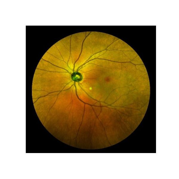
| 6 |  | Laser multicolor image of fundus (Multicolor) | Laser multicolor image has more diagnostic value for a variety of fundus diseases including DR, and it can be used more clearly for image with small pupil and turbidity of dioptric media |
Cases


Advantage
| 1 | More precise | Six image modes: FFA/ICGA/AF/RF/NIR/MULTICOLOR, optical resolution of 5um |
| 2 | More quickly | 1) 100°/60°/30° Wide field view lens optical design, fine imaging 2) Automatic image mosaic, two images can cover the whole retina imaging |
| 3 | More comfortable | 1) Non mydriatic (minimum pupil size as low as 2mm) , can be used for clear imaging of cataract patients 2) No flash/weak light for better experience |
| 4 | More stable | 1) New generation of laser light source, more stable 2) Integrated design, simple and durable |
Specification
| 1 | Model | CRO PLUS |
| 2 | Name | Ultra-wide field laser scanning retina angiograph - CRO PLUS |
| 3 | Imaging mode | FFA + ICGA + AF + RF + NIR + MCOLOR |
| 4 | Optical scanning lens | Optical zoom 100°/60°/30° |
| 5 | Fundus imaging FOV | 160° |
| 6 | Light source | Solid-State LASER |
| 7 | Frame frequency | 16 fps |
| 8 | Fundus tracking | Averaging Real Time Tracking |
| 9 | Minimum pupil size | 2 mm |
| 10 | Lesion resolution | 5 um |
| 11 | Video/image output | Support Avi Jpg Tiff video or image formats |
| 12 | Microclear Unique Technology | Ultra-wide field lens design + real-time fundus tracking + dynamic optical zoom + confocal laser scanning |

|
